Cross Section Of A Bone : Skeleton Anatomy Human Skeletal Stock Vector Colourbox / They are obtained by taking imaginary slices perpendicular to the main axis of organs, vessels, nerves, bones, soft tissue, or even the entire human body.
Cross Section Of A Bone : Skeleton Anatomy Human Skeletal Stock Vector Colourbox / They are obtained by taking imaginary slices perpendicular to the main axis of organs, vessels, nerves, bones, soft tissue, or even the entire human body.. At the outer regions of the section, you can see a dense, thick layer of compact bone. The upper (biting) surfaces of the tooth are at top, with the lower sections (bottom) embedded in the gums and jaw bone (not shown). The outlined area is a cross section of an osteon of compact bone. After a fracture, woven bone forms initially and is gradually replaced by lamellar bone during a process known as bony substitution. Marrow in the shaft of long bones is typically yellow, with red marrow in the head through the cancellous bone.
Diagram with articular cartilage, marrow, spongy bone, medullary cavity, endosteum, diaphysis, and periosteum. At the outer regions of the section, you can see a dense, thick layer of compact bone. The central tubular region of the bone, called the diaphysis, flares outward near the end to form the metaphysis, which contains a largely cancellous, or spongy, interior. And why does the marrow stop where it does, and so sharply? And recall anatomic structures in cross section.
Cross Section Of A Bone Illustration Stock Image C039 1934 Science Photo Library from media.sciencephoto.com Thus, the lamellar pattern as well as the lacunae size differ between trabecular and cortical bone. Compact bone is the outer layer and the spongy bone forms the inner layer. Sketch and label of a cross section of a long bone : Bone markings the surface features of bones vary considerably, depending on the function and location in the body. As the names suggest compact bone looks compact and the spongy bone looks like sponges. The central tubular region of the bone, called the diaphysis, flares outward near the end to form the metaphysis, which contains a largely cancellous, or spongy, interior. Each bone in your body is made up of three main types of bone material: Diagram with articular cartilage, marrow, spongy bone, medullary cavity, endosteum, diaphysis, and periosteum.
Marrow in the shaft of long bones is typically yellow, with red marrow in the head through the cancellous bone.
The surface features of bones vary considerably, depending on the function and location in the body. (area/long bone length 3) ∗ 10 8. Cross section of mandible at first molar region showing cortical and spongy bone basic concepts in osteogenesis bone is a dynamic biological tissue, composed of various metabolically active cells that are integrated into a rigid framework. Related posts of cross section of human bone diagram bone in arm pictures. Internal structure of a human long bone, with a magnified cross section of the interior. While it is not as hard as compact bone, spongy bone plays an important role of protecting the marrow where blood cells are produced. They are obtained by taking imaginary slices perpendicular to the main axis of organs, vessels, nerves, bones, soft tissue, or even the entire human body. The upper (biting) surfaces of the tooth are at top, with the lower sections (bottom) embedded in the gums and jaw bone (not shown). Marrow in the shaft of long bones is typically yellow, with red marrow in the head through the cancellous bone. At the outer regions of the section, you can see a dense, thick layer of compact bone. Now that you know what bones do, let's take a look at what they're made of and their anatomy. Table 1 describes the bone markings, which are illustrated in (figure 4). Explaned distal and proximal epiphysis.
Learn vocabulary, terms, and more with flashcards, games, and other study tools. Bone markings the surface features of bones vary considerably, depending on the function and location in the body. Über 7 millionen englischsprachige bücher. Chapter 6 bones and skeletal tissues flashcards quizlet.shop the edit of floral dresses, dream jeans and fresh shoes now, and stay tuned for a lot more exciting topshop stuff to come. Would it be a good thing to show the epiphyseal plate?
Pediatric Skeletal Growth from www.fairview.org Smartdraw includes 1000s of professional healthcare and anatomy chart templates that you can modify and make your own. Two types of bone tissues in cross section of a long bone : Diagram with articular cartilage, marrow, spongy bone, medullary cavity, endosteum, diaphysis, and periosteum. While it is not as hard as compact bone, spongy bone plays an important role of protecting the marrow where blood cells are produced. The upper (biting) surfaces of the tooth are at top, with the lower sections (bottom) embedded in the gums and jaw bone (not shown). Explaned distal and proximal epiphysis. After a fracture, woven bone forms initially and is gradually replaced by lamellar bone during a process known as bony substitution. Body size standardization was done, using the following equations:
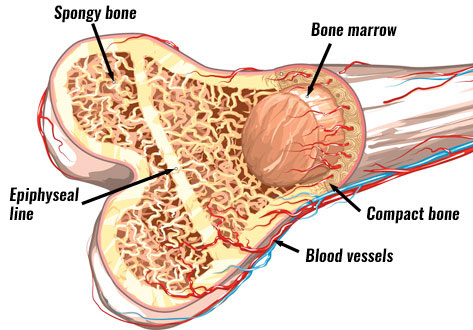
Internal structure of a human long bone.
Now that you know what bones do, let's take a look at what they're made of and their anatomy. Related posts of cross section of a long bone bone test anatomy and physiology. Browse 4,294 bone cross section stock photos and images available, or search for human bone cross section to find more great stock photos and pictures. And why does the marrow stop where it does, and so sharply? Marrow in the shaft of long bones is typically yellow, with red marrow in the head through the cancellous bone. Table 1 describes the bone markings, which are illustrated in (figure 4). Each bone in your body is made up of three main types of bone material: Chapter 6 bones and skeletal tissues flashcards quizlet.shop the edit of floral dresses, dream jeans and fresh shoes now, and stay tuned for a lot more exciting topshop stuff to come. At the end of the bone is the epiphysis, which in young people is separated from the. Concentric layers of bone cells (osteocytes) and bone matrix surround the central canal. Diagram with articular cartilage, marrow, spongy bone, medullary cavity, endosteum, diaphysis, and periosteum. The upper (biting) surfaces of the tooth are at top, with the lower sections (bottom) embedded in the gums and jaw bone (not shown). Would it be a good thing to show the epiphyseal plate?
Would it be a good thing to show the epiphyseal plate? Related posts of cross section of human bone diagram bone in arm pictures. Learn vocabulary, terms, and more with flashcards, games, and other study tools. The central tubular region of the bone, called the diaphysis, flares outward near the end to form the metaphysis, which contains a largely cancellous, or spongy, interior. Vector illustration scheme of bone cross section.
Bone Structure Anatomy Explained What Is Bone Marrow from www.teachpe.com The surface features of bones vary considerably, depending on the function and location in the body. After a fracture, woven bone forms initially and is gradually replaced by lamellar bone during a process known as bony substitution. Wing bones were sampled from the right side of skeletally table 1. Bone markings the surface features of bones vary considerably, depending on the function and location in the body. Internal structure of a human long bone, with a magnified cross section of the interior. Explaned distal and proximal epiphysis. While it is not as hard as compact bone, spongy bone plays an important role of protecting the marrow where blood cells are produced. There are trabeculae in spongy bone which gives its sponge like appearance.
Internal structure of a human long bone, with a magnified cross section of the interior.
Learn vocabulary, terms, and more with flashcards, games, and other study tools. Bone in arm pictures 12 photos of the bone in arm pictures bone cancer arm pictures, pictures of bone cancer in arm, bone, bone cancer arm pictures, pictures of bone cancer in arm Table 1 describes the bone markings, which are illustrated in (figure 4). Diagram with articular cartilage, marrow, spongy bone, medullary cavity, endosteum, diaphysis, and periosteum. Internal structure of a human long bone, with a magnified cross section of the interior. There are trabeculae in spongy bone which gives its sponge like appearance. While it is not as hard as compact bone, spongy bone plays an important role of protecting the marrow where blood cells are produced. Marrow in the shaft of long bones is typically yellow, with red marrow in the head through the cancellous bone. (area/long bone length 3) ∗ 10 8. Wing bones were sampled from the right side of skeletally table 1. The large dark spots are passages for blood vessels and nerves. Internal structure of a human long bone, with a magnified cross section of the interior. In the center of each osteon is the central canal, a space that houses blood vessels and nerves that supply bone.


0 Komentar